CHAPTER 01
Twenty-nine eyes
Twenty-nine eyes, three fragments and three white pieces fit inside a matchbox. The box is covered in blue paper and carries a handwritten German label. It is about six centimeters long. At the Corning Museum of Glass it has an accession number, 93.3.74-37, and a practical description: parts for making marine animals.
Leopold and Rudolf Blaschka kept components like these ready for assembly. An order could arrive from a university or museum, and parts would be selected, joined with wire and glue, then finished and painted. The eyes belonged to a supply of bodies that did not yet exist.
Their completed animals are much better known than the contents of the boxes. They have bells, tentacles, arms, fans, stalks and coils. Some resemble a drop of water that has acquired an elaborate internal arrangement. Others are so thoroughly painted that their material is difficult to recognize. They were made for people who wanted to look closely at a creature and could not conveniently keep it alive.
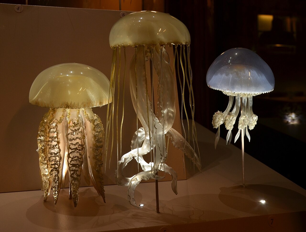
Glass jellyfish models in Geneva’s natural history museum, photographed by Vassil in 2014. Photograph and source record, released under CC0. The display supports are visible; these are models photographed in a case.

Look at the middle model in this photograph. Its upper surface is broad and smooth. Below the rim hang two quite different sorts of structure. Thin strands descend almost vertically around the outside, while wider transparent ribbons twist through the center. The ribbons catch light along their folds. Their edges are easier to follow than their surfaces.
The model on the left is arranged differently. Its hanging parts are crowded with small, repeated frills. Light gathers in the irregularities, giving the lower half a granular appearance. The right-hand model has a bluish bell and a much shorter group of hanging structures. All three share an umbrella-like outline, but a quick name such as “glass jellyfish” loses most of what distinguishes them.
Their shadows are part of the photograph too. There are rods beneath the animals, reflections on the base and small numbered markers. These details help establish what we are looking at. The glass is standing in air. The lighting comes from a display. The animals themselves are absent.
It is possible to admire the models without knowing anything about their makers. It is also possible to become absorbed in their making and forget that the arrangement of each body was intended to say something about an organism. Both approaches miss useful details. A row of tiny knobs might be an anatomical feature, a construction choice or a later repair. A smooth region might be exposed glass, a coating or simply an effect of the light.
This book follows objects and records that let us ask those questions with some precision. The evidence includes photographs, drawings, a workbench, catalog entries, shipping correspondence and the reports of conservators who have examined damaged models. The living animals occasionally intervene by doing something a fixed representation cannot show.
The matchbox offers a useful place to begin because it interrupts the usual view of a finished masterpiece. An eye can be made before there is a head to receive it. A tentacle can wait. Work can be divided into parts, compared, rejected and tried again. The visible delicacy of the final animal does not tell us that it emerged in one uninterrupted performance at a flame.
CHAPTER 02
The specimen in the jar
A jellyfish in a museum jar is an actual jellyfish. A glass model is not. That distinction remains important even when the model looks more alive.
The jar contains biological material. Depending on its history and condition, a researcher may be able to examine structures, take measurements or obtain other evidence from it. The specimen also has a collecting history: a place, a date, a collector, perhaps notes made before preservation. Its value does not disappear because it is difficult to display attractively.
But a visitor looking through the jar may see an indistinct mass. The Natural History Museum in London describes the problem that made the Blaschkas’ work useful: soft marine animals could lose their expanded form and color after preservation. Its glass models drew on scientific illustrations and animals kept in aquaria. They supplied an appearance that the preserved material no longer conveyed well.
A drawing solves some of the same problems. It can separate overlapping parts, enlarge a small structure and show an animal in an informative position. It can also place several views on the same sheet. A model adds depth. You can move your head and see one part pass in front of another. A projection that looked like a line becomes a tube. Two structures that overlapped separate as your viewpoint changes.
Neither form of representation is automatic. Someone has to decide what to make visible. A drawing viewed from one side leaves the other side unspecified. A model supplies that other side, but supplying it requires evidence or judgment. Its solidity can make the judgment harder to notice.
Think of a photograph of a flower taken directly from above. It may show the number and shape of the petals quite clearly while revealing little about their attachment. A side view answers another set of questions. A section through the flower answers others. Making a single object that serves all three purposes could require an enlarged or opened-out model rather than a faithful exterior copy.
Nineteenth-century teaching collections used several materials to solve these problems. In their research on Australian and New Zealand collections, Jan Brazier and Molly Duggins describe wax, papier-mâché, glass models and large wall charts used alongside specimens. The Ziegler workshop made embryological models; Brendel supplied botanical models. A room equipped for teaching could contain several kinds of representation, each doing a different job.
Wax could carry an enlarged developmental stage. A wall chart could make a complicated arrangement visible from the back of a lecture room. Glass could suggest the translucency of a soft animal. These were choices about the lesson as well as choices about the material.
There is a practical test for a teaching model: what can a student do with it? Naming it is only one possibility. The student might trace a channel, compare two stages, count repeated structures or explain how one part attaches to another. An attractive object that does not help with the intended question may be less useful than an awkward-looking one that does.
The Blaschkas’ animals invite a slightly different test now. Many visitors encounter them as valuable historical objects, protected from handling and separated from the lessons for which they were purchased. We can recover some of their teaching use by looking for the question built into the form. Why this scale? Why this position? Why two versions on the same base?
A jar, a drawing and a model can all belong beside one another. The glass makes a body legible. The specimen keeps evidence that no amount of skilled glassworking can replace.
CHAPTER 03
Gosse’s crowded rock
Philip Henry Gosse wanted a reader to be able to distinguish one sea anemone from another. In the preface to his 1860 Actinologia Britannica, he complained about descriptions that were too vague to be useful. He was equally suspicious of excessive detail that merely recorded the peculiarities of one individual.
His solution was orderly comparison. Describe the same kinds of feature in the same sequence, and the reader can put two accounts beside each other. What is distinctive becomes easier to find. Gosse’s book combined that disciplined purpose with colored plates that seem, at first, much less orderly.
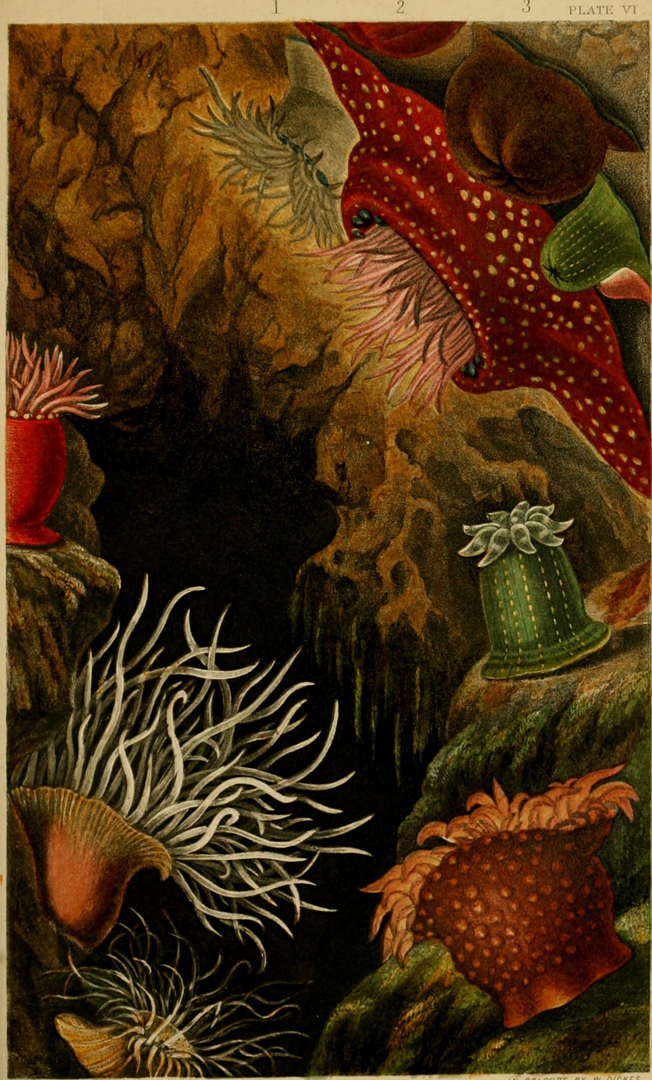
Plate VI from Philip Henry Gosse’s Actinologia Britannica (1860), printed in color by William Dickes from Gosse’s drawings. Public-domain scan from the Smithsonian Libraries through the Biodiversity Heritage Library. Image record; the book.

The plate is crowded. A rough brown rock divides the picture. At the upper right, a red body covered with pale spots occupies a large patch of the page. Pink tentacles project from beneath an overhanging edge. Nearby is a rounded brown animal with its tentacles withdrawn. Lower down, a green column carries short, pale projections around its top. At the bottom left, long white tentacles cross one another in front of a striped body.
The image offers several appearances of anemones rather than one standard silhouette. Expansion and contraction alter how much of the animal is visible. A closed anemone presents a very different identification problem from the same kind of animal fully extended. The rock gives the bodies places to attach and turns the plate into something more spatial than a line of isolated specimens.
Yet the numbers across the top remind us that this is an organized illustration. It is not evidence that every pictured animal occupied one rock at one moment. The composition collects views for the reader. Each numbered figure can be followed into the text.
Gosse described his original drawings as made from life. William Dickes printed the plates in color, trying to reproduce those drawings closely. That adds another person and another process between the animal and the page. Looking at the print, we are seeing Gosse’s selection and drawing, Dickes’s printing and the condition of the surviving copy. Looking at the digital image adds scanning and a screen.
The Blaschkas used Gosse’s book among their sources. The relationship is documented in research on the designs and names in their catalogs. This does not mean that every anemone model is a literal extrusion of one printed figure. It means that a published observation could travel through several skilled hands before becoming a teaching object.
Gosse’s preface is unusually useful for understanding that movement. He explains that he had studied animals from British shores and received specimens from other naturalists. He distinguishes his direct acquaintance with living animals from information obtained through previous books. The work assembled contributions from several places, even though his name stood on its title page.
He also acknowledges a problem caused by publication in installments. Nearly two years had passed while the book appeared in parts. Later sections could correct earlier ones. Readers of the completed volume would therefore find inconsistencies and additions. The physical book preserved those stages of his knowledge together.
A glass model based on a printed account may preserve one such stage too. Its form can remain stable while the name, interpretation or source description changes. That is one reason the paper record matters. If we know which illustration was used, we can ask what the maker could actually have seen there.
Try following one tentacle in the plate with your eye. It may disappear behind another, become difficult to separate from the rock or leave the visible edge of the picture. Now imagine being required to make its complete three-dimensional form. You would need another view, an actual specimen, knowledge of comparable animals or a decision about the missing part.
This small exercise makes the work of model-making more concrete. Copying an illustration into glass is not just changing the material. It requires decisions about depth, attachment and the unseen surface. The quality of those decisions depends on observation as well as dexterity.
The old plate can still teach that distinction. Its color draws us in; its numbers send us back to the descriptions.
CHAPTER 04
An order by number
Cornell acquired its major collection of Blaschka models in 1885 through Ward’s Natural Science Establishment in Rochester, New York. Henry Ward’s business provided a route between a workshop near Dresden and an American classroom. The Corning Museum’s account of the trade places the animals in a catalog business whose offerings had grown to about seven hundred by the late 1880s.
Ordering by number is ordinary until the thing being ordered is a delicate jellyfish with many separate parts. A number makes the transaction manageable. The buyer can specify an item without writing a fresh anatomical description. The seller can connect that request to a design, a price and a set of components.
It does not follow that every object made to the same number is identical. Handwork permits variation. Materials change. A maker may improve an attachment or simplify a base. A collection assembled over several years can therefore hold evidence of working methods as well as a series of organisms.
The number also does not tell us how many pieces should arrive. A single offering may include a mature animal, a juvenile form and enlarged details. If those pieces become separated in storage, each can begin a new life as an apparently independent object. A later curator must work backward from what survives.
A teaching collection has reasons to contain repetition. Two views of one species may answer different questions. A dissection can be useful even when the complete exterior is already represented. A sequence of developmental stages is valuable precisely because the stages differ. Counting the objects without understanding these relationships gives a misleading account of what the collection contains.
The practical paperwork becomes easier to appreciate if you make a small imaginary order yourself. Suppose you want three teaching sets: an adult animal, an adult with an enlarged detail, and a developmental sequence containing four pieces. You have ordered three catalog items and may receive seven physical elements. If a later inventory counts species, the answer could be three, two or one, depending on what the sets represent. None of those counts can be inferred from “seven objects” alone.
The example is invented; the cataloging problem is real. Museum records need to distinguish an individual object from the numbered offering, the biological subject and the collection in which it is now kept.
An old label can help hold those distinctions together. It may include a model number, a name and a reference to the source used for the design. Even a worn card beneath a beautiful model can contain information that the glass itself cannot supply. Removing the card to make a cleaner display may also remove the easiest route back to the original description.
The models traveled through institutions after their first sale. University College London’s Grant Museum records that part of its collection arrived when the Science Museum distributed glass models in the 1920s. Other models were already listed in a Grant Museum catalog compiled in 1890. The present collection brings together purchases with different histories.
Between Dresden and its present case, a model could pass through several collections. Teaching, storage, transfers and repairs each added to its history. A display label has room for only a small part of it.
For a reader following the history, the accession number is often the most useful thing to copy down. Names change; a stable object identifier gives you something to search for again.
CHAPTER 05
Under the table
Rudolf Blaschka’s workbench was built by a family of organ makers. That fact becomes less surprising when you look beneath the tabletop.
The bench has bellows. Moving the treadle drives air from a lower chamber into an upper chamber, which helps supply a steadier flow to the flame. The Corning object record identifies the makers as the Prediger family and dates Leopold’s commission for Rudolf to 1876. Wood, metal and leather form the apparatus. Part of its air-delivery system is now missing.
This was a compact working installation, roughly eighty-five centimeters across the tabletop and about a meter deep. The glassworker’s hands could manipulate the material while a foot helped maintain the flame. The fuel could be paraffin or alcohol. The bellows supplied air; they were not a modern oxygen cylinder hidden in a Victorian table.
The division between foot and hands is easy to overlook in a photograph of the upper surface. A steady flame gives the hands a more predictable material to work with. If the heat changes abruptly, the section being shaped may become too stiff or too soft at the wrong moment. A mechanism that smooths the air supply therefore supports the precision visible in the finished object.
Lampworking begins with glass rods or tubes softened in a flame. The worker can pull, bend, join or blow the heated material. A rod offers solid glass; a tube already contains an opening. Those starting forms are useful for different parts of a model. Neither tells the worker what an anemone should look like.
There are several problems to solve at once. The glass must become workable without losing the form already achieved elsewhere. A thin region heats differently from a thicker one. A part that looks balanced when supported by a tool must eventually stand, hang or attach to something. The maker has to think beyond the moment when the glass is soft.
Consider a long, thin projection. If it is made as part of the whole animal, a mistake may endanger work already completed. If it is made separately, it can be inspected before attachment, but the joint introduces another problem. The maker must decide where to place that joint and how to make it carry the load without obscuring the anatomy.
The surviving stock of parts shows that the Blaschkas often chose separate manufacture and later assembly. It does not make the work less demanding. It moves some of the difficulty from forming the entire animal at once to preparing compatible pieces and joining them accurately.
There is a temptation to describe every fine detail as a feat of hot glass. A close examination can disprove that description. Conservator Astrid van Giffen reported that the small dots on a model of Carinaria were attached with glue. Where a dot was missing, the exposed glass also revealed the surrounding matte coating. What looked like one continuous surface contained several materials and operations.
The finished model conceals much of its order of assembly. A beginner trying to reproduce the visible object in one piece may therefore attempt a harder process than the original makers used. Studying the joins can be more informative than staring at the most impressive contour.
The bench places the seated maker in a particular arrangement: a foot operating the air supply and two hands near a localized flame. The mechanism has to let those activities continue together.
CHAPTER 06
Why the edge lights up
A clear drinking glass can almost disappear against one background and become conspicuous against another. Its edge may shine while its center seems empty. Move a lamp and the bright line moves too.
The Blaschka models use this behavior extensively. In the photograph of the three jellyfish, some thin surfaces are visible mainly because their edges redirect light toward the camera. Others reveal what lies behind them. A translucent bell spreads the light more diffusely and gives a stronger impression of volume.
Transparency and translucency are related but different. Through a transparent region we can retain a relatively clear view of what is beyond it. A translucent region lets light through while scattering it enough to blur that view. A painted or opaque region may prevent us from seeing through it at all. A single model can contain all three.
Glass also reflects light at its surfaces. A highlight can show the position of a lamp or a bright part of the room. It can obscure a small anatomical feature just as easily as it can reveal a curve. Photographing a model is therefore an additional act of description, with its own decisions about what to emphasize.
Corning photographer Andy Fortune explains this directly in his account of photographing the Blaschka collection. For a model of a tube worm, he deliberately used dappled lighting to suggest an underwater setting. He still wanted the result to be recognizable as glass. The appearance was produced through photographic choices, not by submerging the historical object in its animal’s habitat.
That distinction matters when a photograph is detached from its caption. A dark background can make a supporting base disappear. A carefully placed light can make an opaque region glow. The image may be accurate as a photograph of the object while encouraging the viewer to imagine a living animal suspended in water.
You can explore these effects with an ordinary, robust drinking glass on a stable table. Use room light or a small cool lamp. Put white paper behind it, then a dark sheet, and observe which boundaries become easier to see. Move your own viewpoint slightly. Look for the reflected light source. You do not need to heat, coat or alter the glass.
Try placing a sheet with a thick straight line behind the glass. Where does the line seem displaced or distorted? Refraction changes the path of light as it passes between air and glass. The surfaces it crosses have different orientations. Curvature changes those orientations across the object. The line gives you something definite to follow through that distortion.
Now use an empty section of the sheet. The distortion remains, but it is harder to notice because there is no strong feature behind the glass. This helps explain why a model can be difficult to document. A camera needs evidence of a surface, and a clear surface may mostly show the background through it.
The drinking-glass exercise helps us notice light and surfaces; it cannot measure the Blaschkas’ materials. Their models contain different compositions, coatings, thicknesses and internal supports. We cannot identify those materials by comparing them casually with a drinking glass.
Glass at ordinary conditions is a rigid, noncrystalline solid. Heating changes how readily it can deform. Its composition influences its working behavior and optical properties. Calling every glass part the same material misses differences that a maker or conservator may find crucial.
In the jellyfish photograph, follow one of the broad central ribbons from its bright edge into its nearly invisible middle. The form is continuous. The visibility is not. Much of the model’s apparent delicacy comes from that uneven distribution of light.
CHAPTER 07
The blue animal upside down
The glass Glaucus has a silvery body and blue projections arranged in clusters. The largest clusters spread out near the head. Smaller ones follow down the body, and a long tapering tail extends behind. Against the dark background, the individual projections look like fine blue strokes with clear tips.
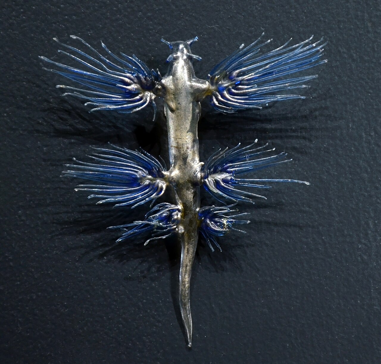
The Geneva model identified as Glaucus atlanticus. Photograph by Vassil, 2014, CC0. Image record. The photograph shows the object’s appearance under display lighting, not a measured record of the living animal’s color.

This is a sea slug. The elaborate silhouette can make that ordinary name seem inadequate, but it belongs to the same broad molluscan world as less spectacular slugs and snails. Its projections are called cerata. The living animal floats at the sea surface, upside down relative to its usual anatomical orientation.
Bill Rudman’s Australian Museum account explains the coloring in relation to that position. The blue side faces upward, while the more silvery side faces down. The animal feeds on floating prey including Physalia, the Portuguese man-of-war or bluebottle, and retains stinging capsules in sacs at the tips of its cerata. A gas bubble helps support its surface life. The attractive stranded animal should not be treated as a harmless object to pick up.
These facts change the way we read the model. The blue projections are not fins attached to a miniature dragon. The animal’s orientation in the water differs from the orientation we may assume when it is laid against a flat background. A photograph can make the head-to-tail axis clear while leaving “up” ambiguous.
There is also a difference between the biological feature and the way the model expresses it. A row of glass projections can indicate arrangement and relative length. It cannot demonstrate their movement, the animal’s buoyancy or the behavior of stinging capsules. The model gives a student something to inspect while an explanation supplies processes that remain invisible.
Look closely at the left and right clusters. They are broadly paired, but the visible strands do not form perfect mirror images. Some overlap; some point at slightly different angles; some catch more light. We cannot determine from this photograph how much of that difference comes from the original making, subsequent damage or the viewpoint. We can record the asymmetry without inventing its cause.
A fine transverse line is visible across part of the body. It would be tempting to call it a repaired break. That would be an interpretation, not an observation established by the picture. A condition report or closer examination might settle the matter. Until then, “a line is visible” is the accurate description.
Age and handwork offer plausible explanations for an irregularity. Deciding between them takes more evidence than the irregularity itself.
The image is already rich without those additions. Follow the blue from a dense cluster near the body toward a clear tip. Notice how the silvery surface catches a broad highlight while the slender projections catch narrow ones. The maker has represented several kinds of visual texture within a small animal.
Then return to the living slug’s orientation. The sea surface is above it; the open water is below. The model’s flat presentation has become a body occupying a particular place in the ocean.
CHAPTER 08
The squid laid flat
The squid’s long structures make loops across the display surface. Several shorter arms point forward. One eye is visible as a small amber center surrounded by blue. At the rear, a pale fin rises from the tapering body.
The photograph identifies this Geneva model as Chiroteuthis veranyi. It is a useful example of how much a model can show and how much its presentation can leave unresolved.
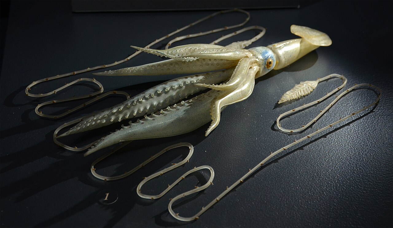
Blaschka model identified as Chiroteuthis veranyi, Geneva. Photograph by Vassil, 2014, CC0. Image record.

The loops let a long part fit within a limited display area. They also make the part’s length more difficult to judge at a glance. A straight measurement from one end of the photograph to the other would not give its full length. You would have to follow the curve, and you would still need a scale.
Some arms overlap. Along their nearer surfaces, repeated raised elements can be seen clearly. Farther back, a bright reflection obscures detail. The visible eye is strongly colored, while much of the body is pale and partly translucent. These contrasts help the viewer separate structures that might otherwise merge into one reflective surface.
The model is not demonstrating a swimming posture. It is resting on a support. We should not convert the arrangement of every loop into a behavioral claim about the living species. A natural-history representation may be positioned for stability, visibility or storage rather than for a literal recreation of the animal in motion.
Modern observations of related squid show how much behavior can add to a static outline. At the Monterey Bay Aquarium Research Institute, Ben Burford and colleagues examined video of juvenile Chiroteuthis calyx. The juveniles have long tails and adopt shapes and orientations resembling Nanomia siphonophores. Their research proposed a defensive mimicry function. Older animals lose the elaborate juvenile tail. This concerns C. calyx, a different species from the model pictured here.
For the pictured species, the question remains open here. The observations of its relative suggest what footage could reveal; they cannot supply the missing observations.
Video also supplies a different kind of evidence. The researcher can see a posture persist, change or recur under different conditions. A sequence can show whether a structure is held rigidly or moves with the rest of the body. Repeated observations can distinguish an occasional position from a common one. The single model cannot do those things.
Its advantage lies elsewhere. It holds an arrangement still long enough for comparison. The eye does not turn away. The arms remain available to count. A student can revisit the same detail without waiting for a living animal to cooperate. For anatomy, that patience is useful.
The two kinds of evidence can be used together. First inspect the fixed form. Then watch footage of an identified living animal and see which assumptions fail. A part that looked decorative may move independently. A long extension may be deployed much farther than the display suggested. A body that seemed substantial in glass may be nearly transparent in water.
The photograph rewards a final look along the nearest arm. Small repeated details remain distinct even where the whole object seems pale and reflective. Making the body recognizable required the maker to manage both scales: the overall squid and the tiny structures that keep it from being a generic squid-shaped ornament.
CHAPTER 09
The fifth bundle of letters
The Australian Museum had glass marine models, but the evidence connecting them to the Blaschkas was not easy to find. A register described them as coming from the museum’s “Old Collection.” That label preserved a place in an inventory without explaining where the objects had originally come from.
In 2009, archivist Patricia Egan investigated. Her account of the search describes a route through trustee minutes, outward correspondence and bundles of incoming letters. The 1879 minutes referred to purchases through a supplier in Prague, Mr. Frič. An outgoing letter asked for Blaschka models. In the fifth bundle of correspondence Egan found letters about the purchases. Three shipments included glass models alongside other specimens and teaching materials. The supplier later asked about the condition of wax models that had crossed the equator in lead chests.
The episode is satisfying because the answer emerges from an administrative trail. It does not require an undiscovered signature scratched into the glass. The models had been acquired as part of a broader effort to equip a museum, and the evidence remained among records of that work.
The absence of an entry in one register did not mean the purchase had not happened. It meant that the first place searched did not supply the needed information. A different category of record preserved the transaction.
That distinction is common in archival work and easy to lose in retelling. “No record found” is a statement about a search. “There was no record” is a stronger claim. “The event did not happen” is stronger again. Egan’s account shows why moving too quickly through those steps can produce a false history.
The shipments also complicate an image of isolated treasures traveling alone. Models made from glass, wax and papier-mâché could belong to one educational purchase. Their materials created different concerns during transport. A wax object crossing warm regions presented a problem unlike the shock sensitivity of a thin glass projection.
The records described by Brazier and Duggins place the Australian purchases within a wider network of suppliers, museums and university teaching. A collection could be obtained for one institution and then lent or transferred for use elsewhere. The teaching purpose helps explain why several materials and kinds of specimen traveled together.
A modern catalog often separates these histories into neat fields: maker, date, material, location. The original paperwork may combine them in less convenient ways. A letter about a crate may mention several objects without listing them individually. A payment can establish that money changed hands without specifying exactly which model was supplied. A later inventory may use a different name.
Reconstructing the history therefore requires matching records, not merely finding one attractive document. The date must fit. The description must fit. The institutional route must be possible. An apparent match can weaken when one of those conditions fails.
This matters for readers using digitized collections too. Search results are shaped by the words entered and the fields that have been indexed. If the model was filed under a supplier’s name, searching only for “Blaschka” may miss it. If the old name differs from the modern one, a species search may overlook the relevant entry.
The Australian Museum’s models were removed from display in 1941. Their later place in an archive did not erase their original teaching purpose. It changed the records and practical arrangements through which people encountered them.
CHAPTER 10
Two names on one label
An old scientific name is not necessarily a mistake. It may have been accepted when the model was made and replaced later. It may also be misspelled, applied too broadly or attached to a form whose identity is now uncertain. Those are different problems.
Eric Callaghan and colleagues examined the names in the Blaschkas’ final catalog in a 2020 taxonomic revision. They reported that only 35.3 percent of the names were still in use in the form applied there, while 3.7 percent lacked a known synonym. They also distinguished catalog items from species and physical parts. One offering for Aurelia aurita could contain as many as fourteen elements. The names of illustrators, including Gosse and Haeckel, had sometimes been treated as though they identified the taxonomic authorities.
Those findings do not mean that nearly two-thirds of the models portrayed imaginary animals. A name can change while the represented organism remains recognizable. Nor does an unresolved name establish extinction. It establishes a problem of identification.
A useful label can preserve both the historical name and a current interpretation. The old name helps connect the object to a catalog, drawing or letter. The current name helps a reader find biological research. Removing either can make one route easier while blocking another.
Cornell’s digital record for a beadlet anemone illustrates the arrangement. It lists Actinia equina as the current name and records an older Blaschka name separately. The page also supplies a Blaschka number, a Cornell number and the photograph’s identifier. These identifiers refer to different parts of the object’s history and documentation.
A reader does not need to memorize all of them. It is enough to recognize that they answer different questions. Which design was offered by the workshop? Which object is held by this institution? Which digital image am I viewing? A single name cannot reliably do all three jobs.
The distinction becomes especially important when comparing photographs. Two institutions may hold versions of the same model design. Two photographs may show one object before and after conservation. A cropped image may hide the base that would have made the relationship clear. Similarity alone does not establish identity.
The Harvard Museum of Comparative Zoology’s record for a sea gooseberry model makes another useful distinction. It connects the object to Ward’s 1878 catalog while indicating that precise dates and locality information are not available in the usual way. A model’s database record can resemble a specimen record without supplying a wild collecting location.
That is sensible, but it asks the reader to pay attention. A manufacturing date is not the date an animal was collected. A museum’s address is not a biological locality. A name in a database can describe an object’s subject rather than material taken from that species.
The same care helps when searching current taxonomic resources. A historical synonym can lead to an accepted name, but the connection may have qualifications. A doubtful identification should remain doubtful when repeated. Copying a database result without its status can turn a provisional association into an apparently settled fact.
The model remains available through these changes. Its label can be updated without altering the glass. That separation is useful: the object preserves an older representation, while the interpretation can improve as research continues.
When both names are visible, the label records a little of that work instead of hiding it.
CHAPTER 11
The commission for plants
In 1886, Harvard botanist George Lincoln Goodale commissioned the Blaschkas to make glass models of plants. Elizabeth Ware and her daughter Mary Lee Ware financed the project. By 1890, an exclusive agreement devoted the workshop’s work to Harvard’s botanical collection.
The Harvard archive’s history records the change. Rudolf traveled to the United States and Jamaica in 1892 to study plants, draw them and collect reference material. While in Cambridge, he saw the models displayed at Harvard. Leopold died in 1895, and Rudolf continued the work afterward. The commission lasted, through renewed arrangements, into the 1930s.
The change of subject did not mean that plant models were a minor decorative addition to a marine collection. It was a sustained undertaking with its own patrons, scientific purposes and working demands. Harvard describes the resulting Ware Collection as about 4,300 models representing 780 plant species. It includes whole plants, enlarged details and other representations needed for botanical study.
A plant offers a different problem from a transparent jellyfish. Much of a leaf is opaque or matte. A hairy surface should not shine like polished glass. A petal may have a delicate gradient of color. A stem has to carry projecting leaves without looking like a metal support disguised as botany.
The familiar phrase “glass flowers” can make the collection sound narrower than it is. Roots, stems, reproductive structures and developmental stages matter as much as blossoms. The representation may need to explain a process or expose an arrangement that cannot be seen on an intact plant.
Harvard’s account of the models’ materials describes an especially revealing detail: the soft-looking surface of common mullein was made with cotton fibers attached to glass. Gelatin could provide raised details such as veins. Those additions supplied textures that the shaped glass alone did not provide.
The visitor’s first question is often whether the object is really glass. A more informative second question is which parts are glass, and what the other materials are doing. A thin support, an adhesive or a surface treatment can make an otherwise impossible arrangement practical.
Botanical models also make scale into an explicit teaching choice. A whole plant can be shown near natural size, while a microscopic structure is enlarged until its parts can be distinguished. The change in scale must be stated. Otherwise the clarity of the model can create a false impression of the size of the living structure.
The exhibition The Blaschkas at the Microscope includes models made between 1889 and 1893 that explain spore-forming plants and fungi. An illustrated Selaginella structure is magnified five hundred times. The enlargement allows a viewer to inspect a relationship that would otherwise require microscopy and preparation.
At that scale, ordinary intuitions become unreliable. A model ten centimeters across at five-hundredfold linear magnification would represent a structure only a fifth of a millimeter across. That calculation is a general illustration, not a measurement of the Harvard object. It shows why a scale label is part of the scientific content.
The commission supported work that could be revisited and refined over years. It also concentrated the output in one institution, unlike the widely distributed marine models. To understand why one collection is scattered around the world while the other is closely identified with Harvard, the contract matters as much as the craft.
Elizabeth and Mary Ware belong in that account because they made the sustained arrangement possible. They commissioned the work, funded it and kept the arrangement going over many years.
CHAPTER 12
A well-made rotten apple
A spot on a glass apple can represent disease. It can also be damage to the model. A conservator must tell the two apart before deciding what to remove.
During preparations for Harvard’s 2017 Rotten Apples exhibition, Scott Fulton described white spotting caused by lead salts on a model’s surface. It resembled part of the depicted disease but was a result of the object’s deterioration. The exhibition report records months of work by Fulton and collection manager Jennifer Brown. The display included seventy-eight glass pieces, with pears, plums and apricots among the apples, and examples of several fruit diseases.
That is a particularly demanding conservation problem. The intended appearance is already irregular. A dark patch may be deliberate. A rough surface may be the information the model was made to preserve. Making the fruit look healthier would make the object less accurate.
It also makes a useful correction to the idea that the Blaschkas selected only pristine specimens. A diseased fruit can be an excellent teaching subject. It shows a process that changes over time and may not be available in a convenient condition when a class needs it.
A fresh specimen continues to change after it is collected. A photograph fixes one view. A model can hold a selected stage in three dimensions, with enough detail to compare it with other examples. The fact that it does not rot further is part of its usefulness.
Harvard’s later Fruits in Decay exhibition extended the focus to blight, rot and disease on several summer fruits. These were historical models brought into a new exhibition arrangement, not newly diseased objects. The distinction between the condition depicted and the condition of the depiction remained essential.
Consider how you might describe such a model without its label. “A brown apple” says very little. “A pale circular patch surrounded by darker tissue” begins to locate a feature. “Several raised spots concentrated on one side” records an arrangement. A diagnosis would require more than those observations, but the observations can be checked by another viewer.
The difference is familiar in plant pathology and medicine: an appearance is evidence, not automatically a diagnosis. In a model, there is an additional layer. The maker intended to represent a particular condition, and the surviving object may have acquired changes of its own. A label, an old photograph and a conservation record can help distinguish those layers.
The glass introduces its own time scale. The represented apple has stopped at one stage of decay. The object has not stopped aging. Coatings can fade, adhesives can weaken and surfaces can change. A permanent representation still requires care if it is to preserve the information for which it was made.
That care sometimes involves leaving an unattractive feature alone. A conservator cannot use “cleaner” as a synonym for “more faithful.” The reference has to be the object’s intended appearance, supported by evidence, rather than a general preference for smoothness or brightness.
There is something exacting about spending so much skill on an apple that looks unfit for a fruit bowl. A particular condition becomes available for sustained attention, including its least attractive features.
CHAPTER 13
Water inside the polyp
The little droplets were on the wrong side of the glass.
In N. Astrid R. van Giffen’s conservation study of model number 173, representing Podocoryne carnea, moisture had accumulated inside hollow polyps. Some parts consisted of one glass bubble inside another. The enclosed space had developed conditions that encouraged deterioration. Because the interior could not be reached, the droplets were left untreated. The report compares the Cornell model, dating from about 1885, with a later Wisconsin example and documents differences in construction. Objects ordered under the same number did not necessarily have identical joins or bases.
The exterior might be accessible to a conservator’s tools while the damaged interior remained out of reach. Seeing through a wall does not make the wall removable. Transparency can make this kind of damage unusually frustrating: it permits inspection while preventing access.
We often think of glass as chemically indifferent to its surroundings. A drinking glass can stand in water without visibly changing during a meal. That familiar experience is a poor guide to every glass composition over a century. According to the Canadian Conservation Institute’s account of glass care, unstable glass may develop a wet-looking surface, deposits or networks of fine cracks, often called weeping and crizzling. These terms describe deterioration, not decorative techniques. Composition and environment both matter.
A sealed glass form makes the environment harder to assess. The air in a gallery is only one part of the problem. A hollow component can contain a smaller, less accessible space with conditions of its own. A photograph that records the outer surface may miss those conditions entirely.
The other materials can also exert forces on the glass. Harvard’s account of the makers and their materials describes internal damage in a tulip-tree leaf model associated with the contraction of organic coatings. The surrounding glass did not have to be struck to fracture. Materials joined successfully in the workshop can age at different rates and respond differently to changes in their environment.
That complicates the seemingly sensible instruction to keep an object as dry as possible. A mixed object may include material that contracts as it dries, coatings that respond to moisture and glass that has already deteriorated. A conservator has to assess the combination. There is no useful household humidity setting that follows merely from the word “Blaschka.”
Even a condition report needs to distinguish visibility from certainty. “Droplets visible inside the upper bulb” identifies an observation. “A crack runs from the join toward the rim” locates another. Explaining the cause requires material evidence, comparison and sometimes monitoring. The record should preserve the observations even if an early explanation later changes.
For a viewer, the practical consequence is modest: a complete-looking model may have problems that are invisible from the gallery floor. Conversely, a visibly incomplete model may have been stabilized successfully. Completeness and stability describe different things.
The untreated droplets in number 173 belong in the published account for that reason. A report of conservation should include what could not be done. Otherwise a later reader might assume that every recorded defect disappeared when the object returned to display.
CHAPTER 14
The repair is another material
A detached tentacle presents two questions before anyone reaches for an adhesive. Where did it belong, and what survives at the join?
The first question is anatomical as well as mechanical. A fragment might fit against several surfaces well enough to be attached. Only one placement may agree with the original arrangement. Earlier repairs can make the problem worse by providing a plausible-looking answer that has already been accepted for decades.
A 2007 report from Museum Wales records tentacles reattached in incorrect positions and old repairs that had discolored or begun to fail. The conservators chose Paraloid B-72 for new joins in that treatment. They wanted a repair that could be removed and that would give way before the thin glass did. Some visibility of the repair also helped distinguish the intervention from the original material. Cleaning required separate investigation because a solution that removed dirt could also remove paint.
A stronger bond can therefore be the worse repair. If the joint survives a force that breaks the neighboring original glass, the repair has transferred the damage. Strength has to be considered in relation to the parts being joined and the stresses they may experience.
Another object can require another choice. At National Museums Scotland, Holly Daws worked on a thick-horned anemone model whose tentacle area had lost substantial glass. Her treatment included a new epoxy support cast using a wax former and silicone mold. Acrylic adhesive attached the support, tentacles and original top. Epoxy was also used to secure running cracks. The museum’s illustrated record makes the added structure and the stages of treatment visible.
These accounts should be read as decisions about particular objects. One team’s rejection of an adhesive for its joins does not turn that material into a universal prohibition. Nor does another team’s successful use make it suitable for every broken glass animal. The condition, proposed role and future removal of a material all enter the decision.
There is also a distinction between attaching an original piece and replacing a missing one. Attachment brings surviving evidence together. Replacement introduces a new interpretation of the gap. A replacement may be necessary to support other parts, to make the object intelligible or both, but its status needs to remain recoverable.
Suppose two tentacles survive on one side and none on the other. Symmetry might suggest the missing arrangement. It would still be an inference. A drawing, another example of the model or an early photograph could strengthen it. An attractive reconstruction alone would not.
The treatment record can carry information that would overwhelm a gallery label: which parts were original, where new material was added, what alternatives were considered and what remains uncertain. Photographs taken during the work may reveal joins that disappear from view after assembly. Without those records, a later researcher could study the replacement as if it were nineteenth-century evidence.
A repaired model can consequently be more informative than an apparently untouched one, provided its history is accessible. The repair has forced someone to examine the parts closely and write down what they found. The result belongs beside the object’s catalog entry, ready for the next person who needs to understand that particular tentacle.
CHAPTER 15
A scan with holes
A transparent object is difficult to turn into a digital solid. The camera sees light passing through it, reflections on it and objects behind it. Those features do not all describe its surface.
Photogrammetry normally works by finding corresponding features in overlapping photographs. A mark that appears in several views helps locate a point in space. With reflective or transparent materials, the apparent mark may move as the viewpoint changes. A bright patch can belong to a reflected lamp rather than to a painted patch on the glass. A background detail can appear through the body without belonging to the body at all.
Christopher Fried and colleagues addressed this problem in a 2020 study of Blaschka models. They combined photographs with computed tomography, which records differences in X-ray attenuation. For a Diazona violacea model, MCZ SC-418, they used 699 photographs. The photographic and CT reconstructions each missed areas that the other captured. Merging their geometry and adding photographic texture produced a more complete result. Cross-polarized lighting helped the photography; surface sprays commonly used to make difficult objects easier to scan were unsuitable for these fragile historical surfaces.
The number of photographs is striking, but it is not a measure of accuracy by itself. Many nearly identical views can leave the same underside unseen. A smaller set with useful coverage may contribute information that another hundred frontal views cannot provide. The arrangement of the views matters.
A digital model also contains several kinds of information that are easy to confuse on screen. Geometry describes the shape: points, edges and surfaces. Texture supplies an appearance mapped onto that shape. Lighting in the viewer then changes how the combination looks. A convincingly shaded surface can conceal an area where the geometry is approximate.
You can see the distinction with a paper cube. Draw a dark circle on one face. The circle changes the appearance without changing the cube’s shape. Cut a hole at the same location and the geometry changes. From a carefully chosen frontal view, the two might look similar. Turning the cube reveals the difference.
The same question is worth asking of a digital museum object. Does a visible opening pass through the geometry, or is it represented in the surface image? Does a delicate branch have thickness? What happens to the underside when you rotate it? These are questions about the record’s construction, not accusations that the record is false.
The Australian Museum provides three Blaschka models for online rotation, including models identified as Phymactis diadema and Aureliana augusta. They offer views that a single photograph cannot. The museum identifies photogrammetry as the method and links the individual records. The controls and availability belong to the museum’s external service, so this edition links to them rather than claiming to hold its own scans.
A useful digital record preserves its method alongside its appearance. It should say what was scanned, when, by whom and with which important limitations. If geometry from one method has been combined with color from another, that is part of the description.
The screen gives you freedom to turn the object without risking the original. It cannot supply an unrecorded surface. When a scan has a hole, the honest answer may be to leave the hole visible until another view or another method provides evidence to fill it.
CHAPTER 16
Making it again
Vincent Desparrois kept small glass pieces in empty Altoids tins while working out how to recreate a Blaschka anemone. Only later did he discover the workshop’s use of matchboxes for comparable storage. In his 2016 account at Corning, the resemblance emerged from the work itself.
His subject was Ulactis muscosa. He separated the model into components that could be made and assessed before assembly, keeping earlier attempts for comparison. Studying the historical object also encouraged him to consider materials other than glass. The account describes practical investigation by a contemporary flameworker, not the discovery of a single secret formula.
That is enough to dispose of the claim that no one today can reproduce the Blaschkas’ work. It does not mean every question about their techniques has been answered. A modern reconstruction can resemble an original while differing in glass composition, heat source, join or sequence of assembly. Those differences are precisely what a careful reconstruction can help expose.
There are several possible goals for making a model again. One is to match the visible appearance. Another is to test a proposed construction sequence. A third is to reproduce the behavior of particular historical materials. Success at one does not automatically establish success at the others.
For instance, an opaque replacement might match the color of a component in a frontal photograph while transmitting light differently from the original. A modern adhesive might make assembly easier without telling us whether the historical join could have been made in the same order. A flexible support might solve a handling problem that the original makers had to solve another way.
None of those choices invalidates the new object. They need to be stated if the object is being used as an experiment. Otherwise the ease of a modern step can quietly become a claim about the historical workshop.
Failed pieces deserve records too. If three proposed methods consistently collapse a thin form and a fourth works, that is useful information. It is still not proof that the Blaschkas used the fourth method. A different glass, tool or practiced movement might change the outcome. Reconstruction narrows possibilities most effectively when it records its conditions.
The tins are useful because they preserve comparison. The newest attempt may feel obviously better while it is in your hand. Set it beside the earlier ones and you can identify what changed: a narrower stalk, a cleaner join, a different curve. Improvement becomes something visible rather than a memory of difficulty.
You can borrow that habit without learning flameworking. Draw the outline of the squid’s arms from the photograph, set the drawing aside and try again after looking at the attachment points. Keep both drawings. The differences show where your first understanding was vague. Neither drawing is a scientific measurement, but the comparison tells you what to inspect next.
The historical boxes and the modern tins also restore an ordinary part of skilled work to the story. Finished objects leave a workshop looking resolved. Their stored components show how much selection took place before anything was ready for the case.
CHAPTER 17
Back into the sea
Drew Harvell and videographer David O. Brown took the models as starting points for a search for living counterparts. The Harvell Lab describes the project as a way to document marine diversity through film and photography. It contributed to Harvell’s A Sea of Glass and Brown’s Fragile Legacy. Those are accounts of their research and fieldwork; the photographs in this book are museum records, not images from their expeditions.
The move from glass to living animal changes the questions. A model has a fixed outline. An animal may contract, feed, turn or disappear into a shelter. A feature that looks decorative in the case may become understandable when it moves. The squid comparison earlier in this book is one example of how video can reveal behavior that a static object cannot contain.
Identification comes first. A vaguely similar animal is not enough. Old names, revised classifications and incomplete labels can lead a search in the wrong direction. Even a widely circulated image can acquire the wrong name: the Harvell Lab corrected a 2016 identification of a glass worm, specifying Pista cretacea rather than Riftia, a deep-sea tube worm.
Finding a living match would establish that the animal occurred at that place and time, with the usual qualifications about identification. Failing to find one would be harder to interpret. Was the search made in suitable habitat? At the right depth or season? Could the animal have been hidden? How much area was examined?
A museum catalog cannot answer those questions about the nineteenth century. It records what was offered for sale, with all the effects of illustration availability, workshop interest, customer demand and practical manufacture. It is not a survey in which every patch of coast received equal attention.
Imagine trying to reconstruct the birds of a town from a shop’s selection of bird ornaments. The selection would certainly tell you something about which birds people recognized or wanted to buy. A shelf with many kingfishers would not establish that kingfishers outnumbered sparrows outdoors. The same caution applies when glass animals are asked to stand in for past abundance.
A useful comparison with the past needs records of the search as well as records of the animals. A dated specimen with a locality can establish a historical occurrence. Repeated surveys using comparable methods may support a claim about changing abundance. A model connected only to an illustration offers a different kind of evidence: a history of representation and scientific attention.
Those limits leave plenty worth doing. A model can prompt a visitor to learn that the represented animal still exists, where it lives and how it behaves. It can direct attention to small or transparent creatures that rarely appear in a general picture of the sea. It can also make a historical scientific name worth following into a modern database.
The living counterpart should be allowed to surprise us. It may look less jewel-like than the model in warm museum light. Its color may depend on illumination and viewing conditions. Its ordinary behavior may be more revealing than its most photogenic pose.
The comparison is most useful when both records retain their circumstances: this glass object, made from these references; this animal, observed here on this date. Then the resemblance can be examined, and so can the differences.
CHAPTER 18
One model, five minutes
Return to the squid photograph. There is no need to remember the entire history of the workshop before looking at it again.
Start at the blue eye. Follow the outline toward the arms, then choose one arm and trace it as far as the photograph allows. Where does it cross another part? Where does the background disappear behind it? Where does the line become too faint to follow? A finger held above the screen can help, without touching a museum case or mistaking a reflection for a piece of glass.
Now look at the support. The black base and the arrangement above it belong to the photograph’s account of the object. Some parts would be difficult to display without support. The mount helps make the animal visible while also fixing an orientation. Ask which of your impressions came from that orientation.
Move to the blue sea slug. Compare the repeated clusters along its body. Count only what you can actually distinguish. If two branches overlap, leave the count uncertain. The purpose is to see how much information is available from this view, including the places where another view would help.
Then read the captions. They name the photographer, the collection context and the source record. Those details let you return to the image later or find it at a different resolution. They also prevent a photograph of a model from drifting into circulation as a photograph of an animal.
You can save a short note with three parts: something visible, something the record establishes and something you still want to know. For the squid, that might be a visible loop of glass, the name Chiroteuthis veranyi in the image record and a question about how the longest pieces attach. The three statements need not be equally certain.
If the model catches your attention, follow one thread. The old name may lead into taxonomy. A visible seam may lead to a conservation report. The animal’s shape may lead to a modern video. The workshop’s bench may lead to the tools and materials of flameworking. There is no required order after the first close look.
These objects reward time because they contain decisions at several scales. A broad silhouette makes the animal recognizable. A small curve locates a structure. A join solves a problem that disappears from sight when the parts are assembled. The museum record may preserve the name of a purchaser or the date of a repair that explains why the object looks as it does now.
The twenty-nine eyes in the matchbox have not yet become a finished animal. That unfinished state makes them unusually helpful. You can see the stock from which a maker would choose, before the chosen piece took its place and the alternatives stayed in the box.
Sources & edition note
This is an AI-generated, source-based account under the fictional editorial pen name Nico Thread. It contains no invented visits, interviews or workshop dialogue. Close readings refer to the photographs reproduced here; hypothetical examples are identified in the text.
The three museum photographs are by Vassil, released under CC0. The 1860 Gosse plate is public domain. Captions link to provenance records. No generated image is presented as a historical object or a living specimen. Museum scans, films and conservation photographs are linked at their owners’ sites.
Historical names and catalog counts retain their stated scope. Conservation reports describe particular treatments; they are not instructions for repairing an object at home. The current Harvard collection page supplies the figure of 780 species; older institutional pages use differing totals. The sources were checked on 5 September 2026.
- Corning · Matchbox 93.3.74-37 ↗
Object record for the twenty-nine eyes and other workshop parts.
- Corning · Rudolf Blaschka’s workbench ↗
Prediger family, 1876; dimensions, materials and air bellows.
- Cornell · Blaschka collection ↗
Institutional collection and digital object records.
- Corning · Fragile Legacy exhibition ↗
Cornell’s 1885 acquisition, Ward’s distribution and catalog business.
- Natural History Museum · Color in the collection ↗
Models, illustrations and the appearance of preserved soft animals.
- Gosse · Actinologia Britannica (1860) ↗
Public-domain original; preface, observational method and plates printed by William Dickes.
- Callaghan and colleagues · Taxonomic revision (2020) ↗
Catalog items, historical names, synonyms and illustrator credits.
- Patricia Egan · Researching the glass models ↗
The archivist’s account of the 1879 records and Frič correspondence.
- Australian Museum · Collection history ↗
The models’ institutional history and removal from display in 1941.
- Brazier and Duggins · Visualising nature (2015) ↗
Primary research on teaching models, charts and institutional transfers.
- Harvard · Blaschka archival records ↗
Commission, Ware funding, exclusivity, travel and correspondence.
- Harvard · Glass Flowers ↗
The Ware Collection; current main page reports 4,300 models and 780 species.
- Harvard · The makers and their materials ↗
Cotton, gelatin, glass, coatings and internal fractures.
- Harvard · Rotten Apples (2017) ↗
Scott Fulton and Jennifer Brown; deterioration versus depicted fruit disease.
- Harvard · Fruits in Decay (2019) ↗
Historical fruit models depicting blight, rot and disease.
- Harvard · The Blaschkas at the Microscope (2024) ↗
Enlarged botanical and fungal structures, including Selaginella at 500×.
- van Giffen · Conservation of model 173 ↗
2017 conference paper, published 2019; enclosed moisture and construction differences.
- van Giffen · Microscopes and glass conservation (2016) ↗
Glued dots and a matte coating on a Carinaria model.
- Museum Wales · Conserving the Blaschka models (2007) ↗
Incorrect old repairs, surface paint and the rationale for selected adhesives.
- Holly Daws · Conserving an anemone ↗
National Museums Scotland treatment record, including an added support.
- Canadian Conservation Institute · Care of ceramics and glass ↗
Unstable glass, weeping and crizzling.
- Fried and colleagues · Digital preservation (2020) ↗
Photogrammetry and CT; complementary reconstructions of Diazona violacea. DOI 10.1016/j.daach.2020.e00147.
- Australian Museum · Blaschka models in 3D ↗
External photogrammetric models and original object records.
- Vincent Desparrois · Recreating Ulactis muscosa (2016) ↗
Contemporary practical study and stored components. Bench airflow is checked against Corning’s object record.
- Andy Fortune · Photographing the collection (2020) ↗
The photographer’s lighting decisions for glass models.
- Harvell Lab · Living Blaschka invertebrates ↗
The project with David O. Brown, A Sea of Glass and Fragile Legacy.
- Harvell Lab · Image identification correction (2016) ↗
The pictured glass worm was Pista cretacea, not Riftia.
- MBARI · Juvenile squid mimicry (2014) ↗
Burford, Robison and Sherlock’s observations of Chiroteuthis calyx, distinct from the pictured C. veranyi.
- Bill Rudman · Glaucus atlanticus (1998) ↗
Australian Museum account of orientation, buoyancy, prey and stinging capsules.
- Corning · Glass, the stone that pours ↗
Glass as a noncrystalline solid and its working behavior.
- OpenStax · Refraction ↗
Basic optics reference; the drinking-glass exercise is original.
- Illustration credit · Photograph and source record ↗
Source image, creator and public-domain or CC0 status. See the in-text caption.
- Illustration credit · Image record ↗
Source image, creator and public-domain or CC0 status. See the in-text caption.
- Object record · distributed glass models in the 1920s ↗
Individual collection record cited in the text.
- Illustration credit · Image record ↗
Source image, creator and public-domain or CC0 status. See the in-text caption.
- Illustration credit · Image record ↗
Source image, creator and public-domain or CC0 status. See the in-text caption.
- Object record · *Actinia equina* ↗
Individual collection record cited in the text.
- Object record · record for a sea gooseberry model ↗
Individual collection record cited in the text.